Automated high content image acquisition and analysis for drug discovery and cell biology

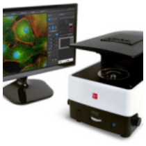
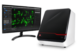
The CELENA® X High Content Imaging System is an integrated imaging system designed for rapid, high content image acquisition and analysis. Customizable imaging protocols, image-based and laser autofocusing modules, and a motorized XYZ stage simplify well plate imaging and slide scanning. The integrated CELENA® X Cell Analyzer software processes images and data for quantitative analysis. Analysis pipelines can be put together and reused to identify cellular or subcellular objects, process images for optimal data collection, and make various measurements. The CELENA® X is as flexible as it is powerful, with interchangeable objectives and filter cubes to accommodate a wide range of fixed and live cell imaging applications.
Features
Fully automated plate and slide imaging
Automated vessel handling and scanning
Motorized XYZ stage, filter cube stage, and objective turret
Laser Autofocus
Rapid and reproducible focusing

Minimized phototoxicity and photobleaching
Live Cell Assay Support
Onstage incubation system for a variety of experiments in physiological and non-physiological conditions
Four Imaging Modes
Fluorescence imaging in four channels, brightfield, color brightfield, and phase contrast imaging
Powerful, Easy-To-Use User Interface
Simple setup of imaging protocols
Seamless integration of imaging and data analysis processes
Customizable High Content Analysis
Create and customize image analysis projects
Quantitatively analyze multiple image-based phenotypes
Create Your Own Imaging And Analysis Workflows With The CELENA
 Imaging
Raw images
Metadata
Image Analysis
Morphology of individual cells and organelles
Spatial distribution of targets
Multiple measurements per cell
Differentiation of multiple phenotypes
Analyzed images
Result files
Graphs (coming soon)
Applications
- Cell Counting
- Cytotoxicity_BF
- Transfection Efficiency
- Wound Healing
- Apoptosis
- Cytotoxicity_FL
- Confluency
- Calcium Signaling
- Phagocytosis
- Cell Cycle
- Stitching
- 3D Cell Models
Versatile And Customizable For Your Cell Imaging Needs
Vessel Holder Selection Guide
A collection of vessel holders are available to accommodate various flasks, dishes, plates, and slides.
Objective Selection Guide
Download our objective selection guide to see the Olympus objectives we recommend for the CELENA® X.LED Filter Cube Selection Guide
Download our LED filter cube selection guide to find which filter cubes fit your needs the best.