Digital Microscope

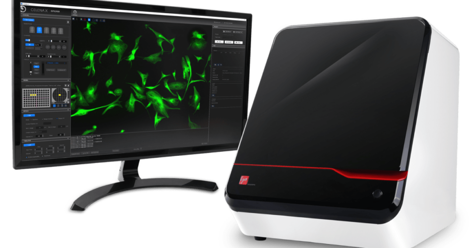
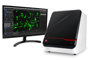
CELENA® S Digital Imaging System
Multicolor fluorescence imaging to data analysis in one device
The CELENA® S Digital Imaging System makes capturing high resolution, publication-quality images a breeze. Don’t let its size fool you, the CELENA® S is equipped with advanced precision optics, a highly sensitive CMOS sensor, digitally controlled LED light sources with hard-coated fluorescence filters, and a computer with image analysis software. The sophisticated yet simple software supports multicolor fluorescence imaging, brightfield imaging, phase contrast imaging, live cell time lapse imaging, and Z-stack imaging.
The All-In-One System for Exceptional Imaging Quality
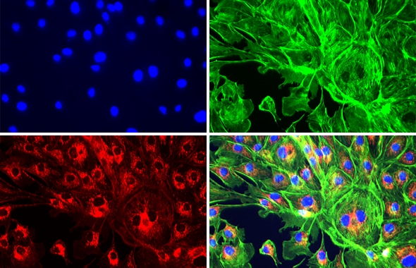
VIBRANT, PUBLICATION-QUALITY IMAGES
Advanced precision optics and a highly sensitive camera allow you to capture high resolution, multi-channel fluorescence images.
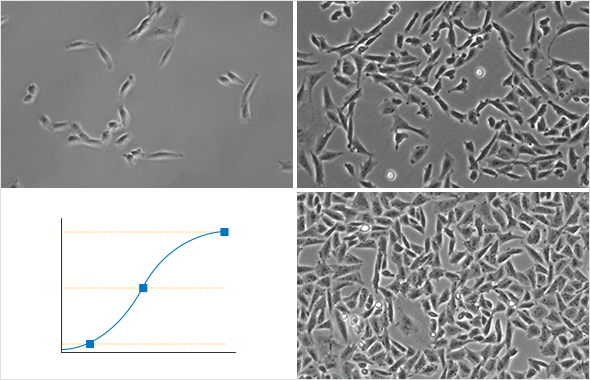
LIVE CELL TIME LAPSE IMAGING
Live cells can be imaged in time lapse mode in a precisely controlled environment. The images can be used to create a video with annotations.
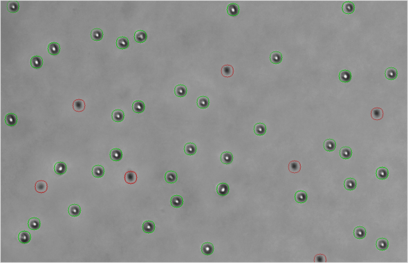
CELL COUNTING
Check cell concentration and viability in cell counter mode. The CELENA® S automatically adjusts light and focus for optimal results.
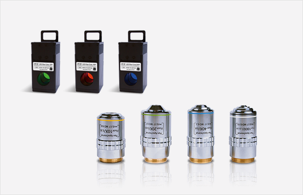
INTERCHANGEABLE FILTER CUBES AND OBJECTIVES
12+ hard-coated LED fluorescence filters. Infinity-corrected objectives with a magnification range of 1.25-100X
LIVE CELL IMAGING MADE SIMPLE WITH AN ONSTAGE INCUBATOR
The CELENA® S onstage incubation system can be set up for a variety of live cell imaging experiments in physiological and non-physiological conditions. The gas mixer allows precise control over the composition, humidity, and temperature of the air. The temperature controller heats the plate and lid of the stage incubator separately, for increased control over the temperature of the system as well as preventing condensation formation.
The CELENA® S makes it easy to set up a time lapse imaging sequence for lengthy cell-based assays, as well as exporting images or creating time-lapse videos complete with annotations.
A collection of vessel holders are available to accommodate various flasks, dishes, plates, and slides.
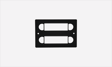
Download vessel holder selection guide
Objective Selection Guide
Our objectives provide excellent resolution, high contrast imaging, and color correction.
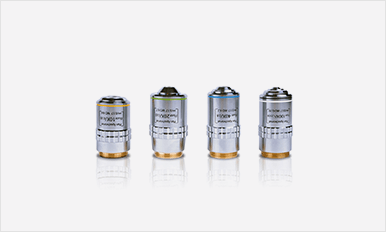
Download lens objective selection guide
LED Filter Cube Selection Guide
Download our LED filter cube selection guide to find which filter cubes fit your needs the best.
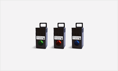
Download LED filter cube selection guide
How to use the CELENA-S
Capturing perfect images
Image analysis
Parfocal correction
Focus memory and fine AF
Z-stack imaging
Focus stacking images

Automated high content image acquisition and analysis for drug discovery and cell biology
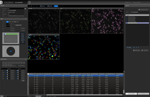
 The CELENA® X High Content Imaging System is an integrated imaging system designed for rapid, high content image acquisition and analysis. Customizable imaging protocols, image-based and laser autofocusing modules, and a motorized XYZ stage simplify well plate imaging and slide scanning. The integrated CELENA® X Cell Analyzer software processes images and data for quantitative analysis. Analysis pipelines can be put together and reused to identify cellular or subcellular objects, process images for optimal data collection, and make various measurements. The CELENA® X is as flexible as it is powerful, with interchangeable objectives and filter cubes to accommodate a wide range of fixed and live cell imaging applications.
Features
Motorized XYZ stage, filter cube stage, and objective turret
Rapid and reproducible focusing
Minimized phototoxicity and photobleaching
Onstage incubation system for a variety of experiments in physiological and non-physiological conditions
Fluorescence imaging in four channels, brightfield, color brightfield, and phase contrast imaging
Simple setup of imaging protocols
Seamless integration of imaging and data analysis processesCustomizable High Content Analysis
Quantitatively analyze multiple image-based phenotypes
Create Your Own Imaging And Analysis Workflows With The CELENA
 Imaging
Raw images
Metadata
Image Analysis
Morphology of individual cells and organelles
Spatial distribution of targets
Multiple measurements per cell
Differentiation of multiple phenotypes
Result files
Graphs (coming soon)
Applications
- Cell Counting
- Cytotoxicity_BF
- Transfection Efficiency
- Wound Healing
- Apoptosis
- Cytotoxicity_FL
- Confluency
- Calcium Signaling
- Phagocytosis
- Cell Cycle
- Stitching
- 3D Cell Models
Versatile And Customizable For Your Cell Imaging Needs
A collection of vessel holders are available to accommodate various flasks, dishes, plates, and slides.
Download our objective selection guide to see the Olympus objectives we recommend for the CELENA® X.
LED Filter Cube Selection Guide
Download our LED filter cube selection guide to find which filter cubes fit your needs the best.
New England BioGroup
NH 03811-1231.
Toll Free USA: 800-779-1016
Main: 617-286-4632
Fax: 617-344-0055
E-mail: info@nebiogroup.com Social: