Skip to content
Products
About Us
News
Contact us
Search
Search
Close this search box.
Search
Search
Close this search box.
Automated Cell Counters
Digital Microscopes
ELISA Automation
Gel Documentation Systems
In Vivo Imaging Systems
Microplate Readers
Microplate Washers
Radio HPLC Detectors
Tissue Dissociators
Tube Luminometers
Tissue Clearing System
Products
About Us
News
Contact us
Automated Cell Counters
Digital Microscopes
ELISA Automation
Gel Documentation Systems
In Vivo Imaging Systems
Microplate Readers
Microplate Washers
Radio HPLC Detectors
Tissue Dissociators
Tube Luminometers
Tissue Clearing System
Our Products
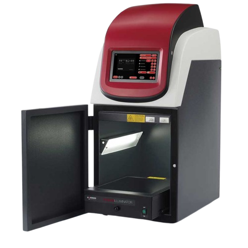
Syngene NuGenius XE
NuGenius XE: Advanced DNA Imaging System for Precise Analysis is a complete, standalone gel documentation...
Details
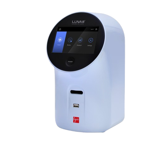
Luna-III | Automated Cell Counter
An advanced brightfield cell counter. Autofocus, 5MP camera, and fast, accurate and reproducible.
Details
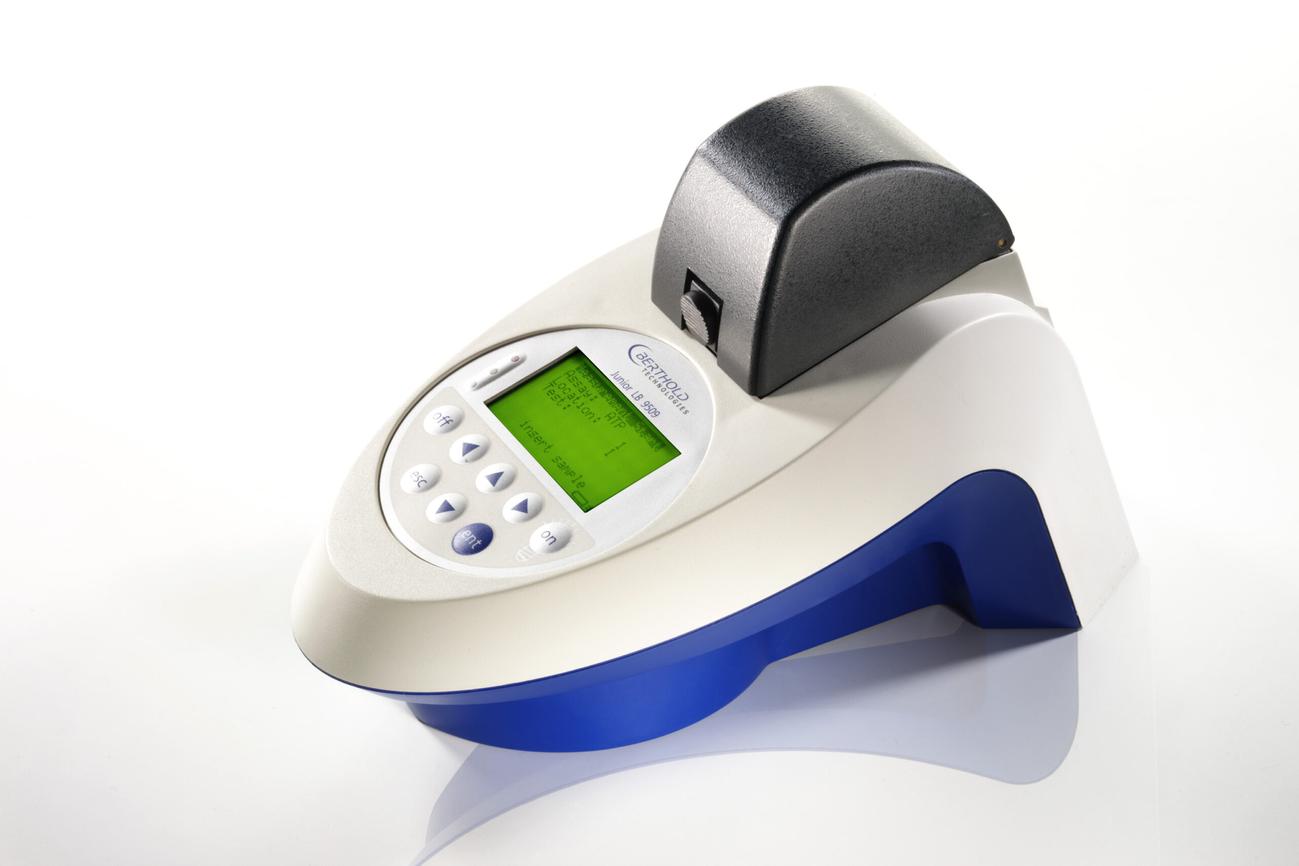
Junior LB 9509 Portable Luminometer
The Junior is a small portable tube luminometer which can be used for all common applications using glow...
Details
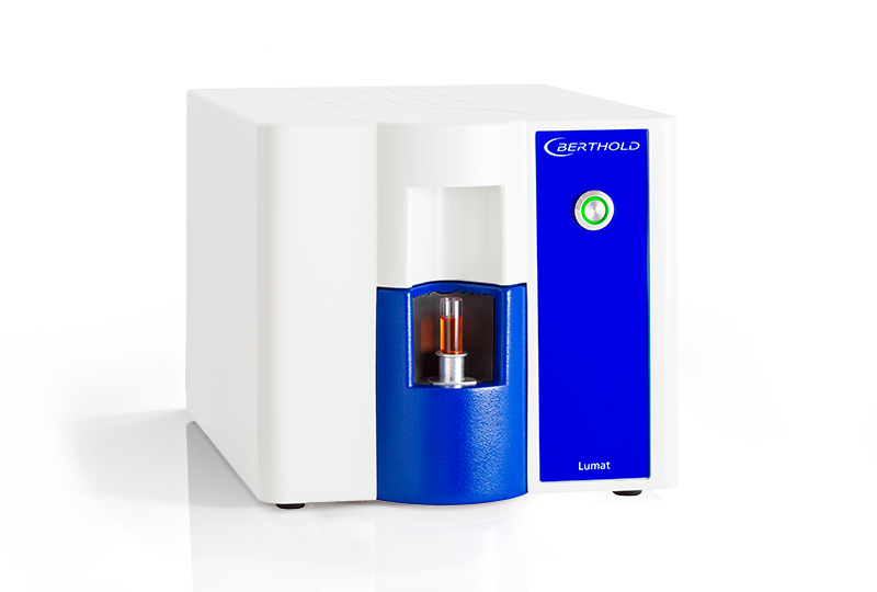
Lumat LB950 Tube Luminometer
The new Lumat is a tube luminometer that uses state-of-the-art technology based on more than 40 years...
Details
NightOWL II LB 983 In Vivo Imaging System
The NightOWL II uses an ultrasensitive back-illuminated (backlit) CCD camera with high quantum efficiency...
Details
NightSHADE evo In Vivo Plant Imaging System
The NightSHADE evo uses an ultrasensitive back-illuminated (backlit) CCD camera with high quantum efficiency...
Details
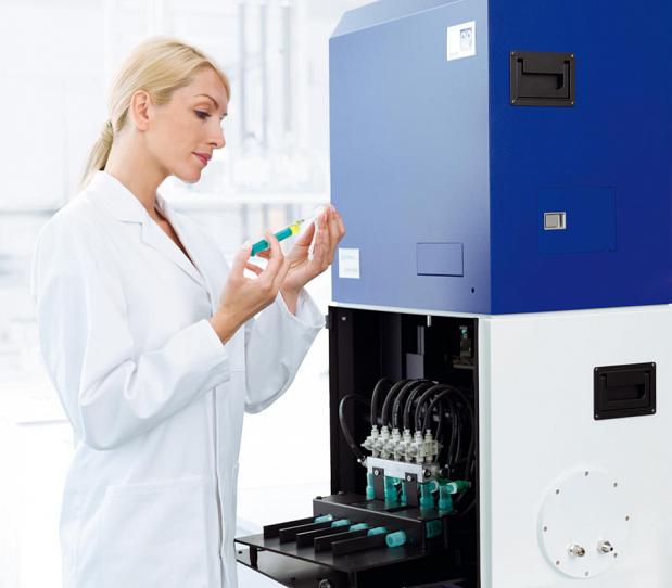
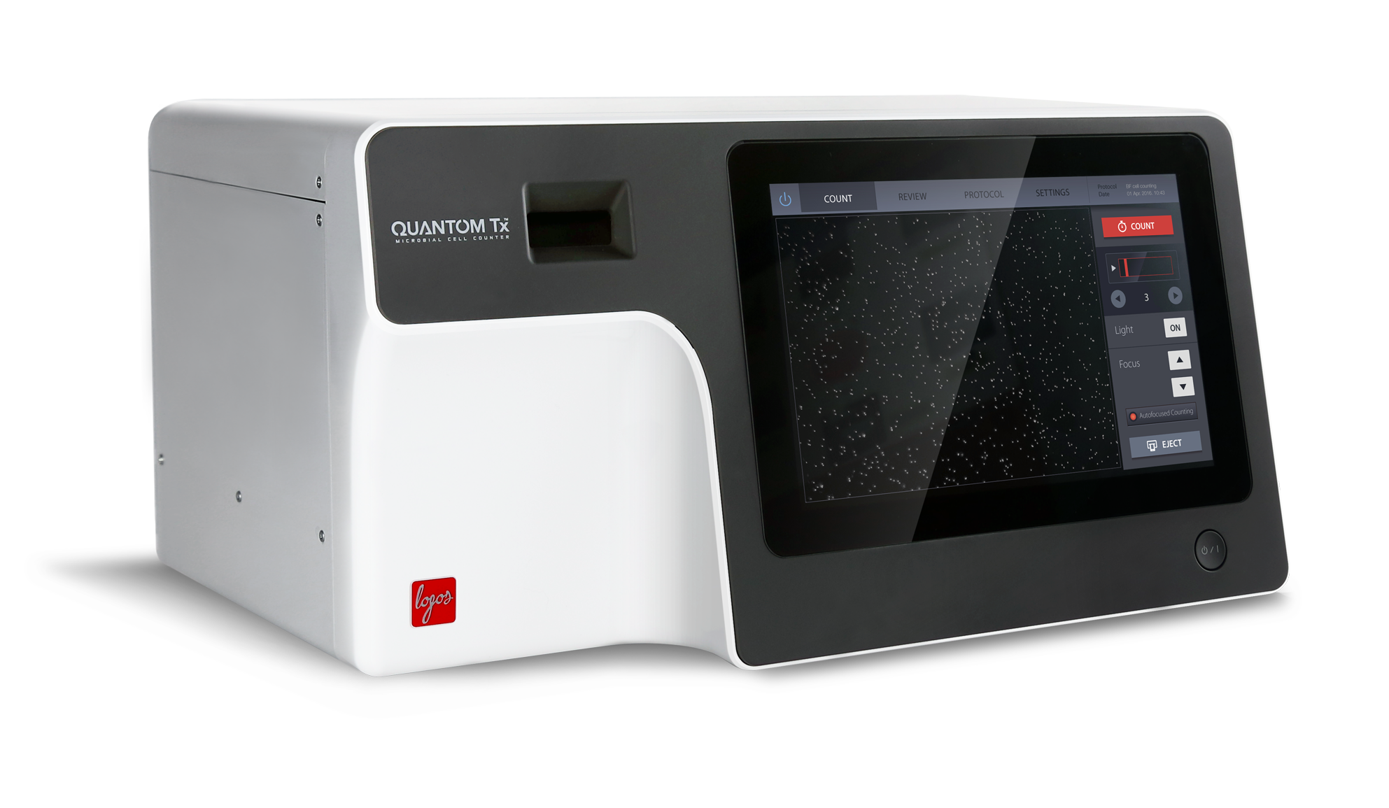
QUANTOM Tx™ Microbial Cell Counter
No more waiting days to count CFUs. In less than 30 seconds, the QUANTOM Tx™ scans up to 10 fields of...
Details
DeepLabel™ Antibody Staining Kit for Cleared Tissues
DeepLabel™ Antibody Staining Kit enhances antibody penetration into large clarified tissues for vibrant...
Details
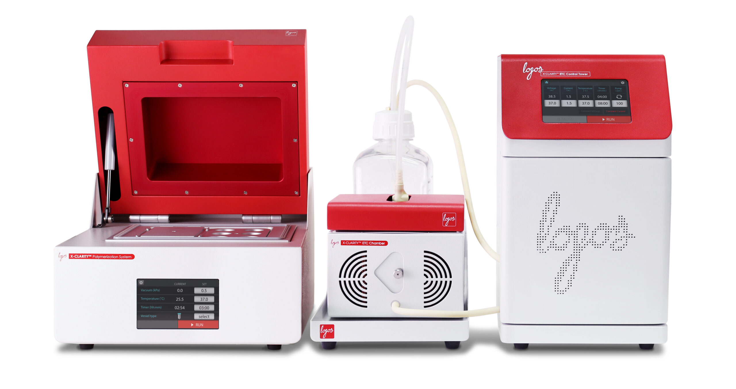
X-CLARITY™ Tissue Clearing Systems & Reagents
The X-CLARITY™ is a collection of systems and ready-to-use reagents developed by Logos Biosystems to...
Details
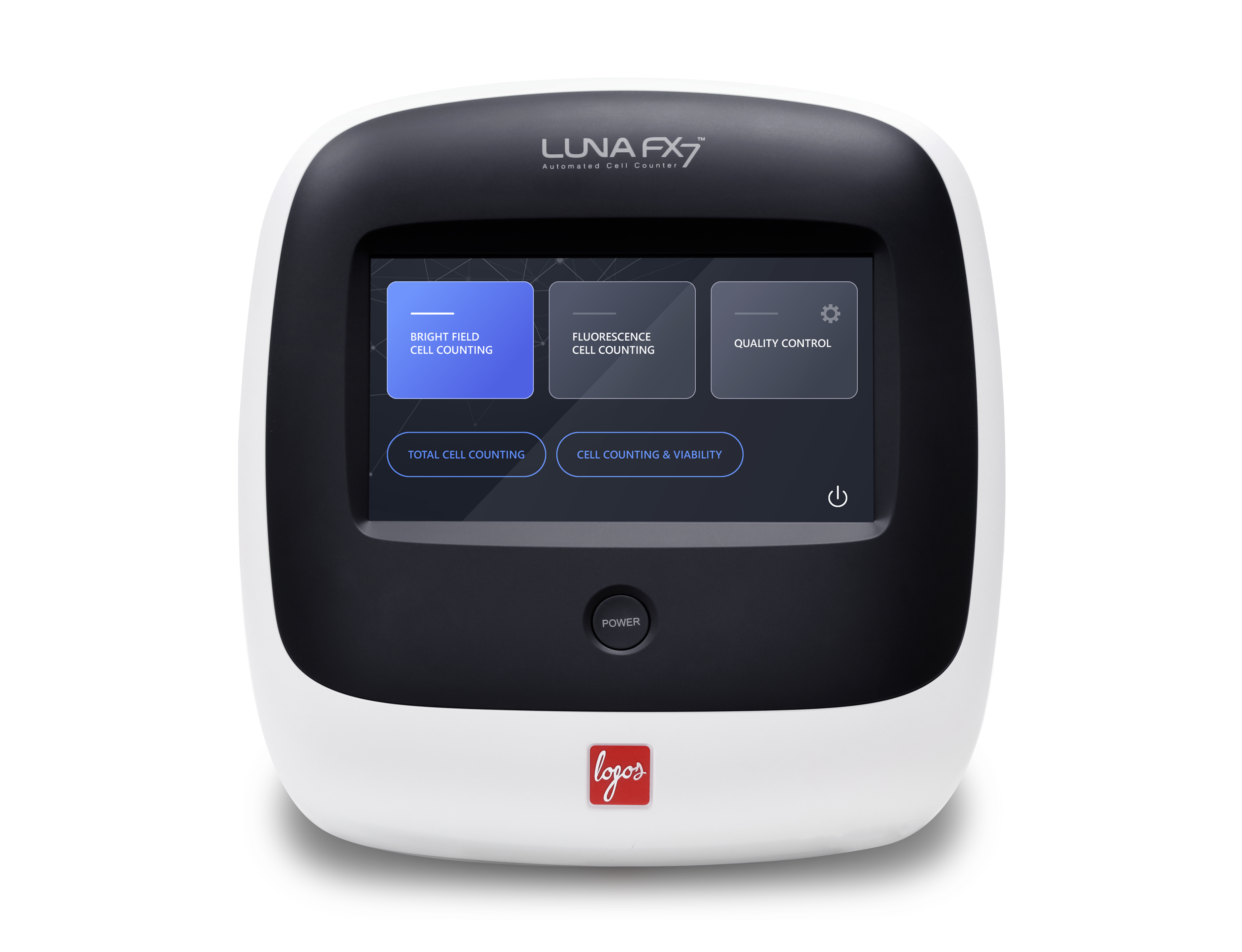
LUNA-FX7 | Automated Cell Counter
The LUNA‑FX7 automated cell counter is a high throughput brightfield and fluorescence cell counter with...
Details
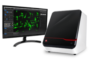
CELENA X|High Content Imaging System
Fully automated plate and slide imaging Automated vessel handling and scanning Motorized XYZ stage, filter...
Details
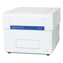
Centro Microplate Luminometer | Microplate Reader
The Centro supports all basic luminescence technologies including: Flash-type Luminescence Glow-type...
Details
No products found
1
2
3
Next